How is lymphatic filariasis spread? Collapsed The disorder distributes to everyone by mosquito bites. When a mosquito bites people who have lymphatic filariasis, microscopic worms are distributed in the consumer’s blood and pollute the mosquito.
What is filariasis?
Filariasis is an infection caused by a parasitic worm and transmitted by insect bites. It can be much more common in the temperate areas of Africa, Asia, and Central and South America. In India, it is prevalent in eastern Uttar Pradesh and Bihar. This disorder spreads to everyone through mosquito bites. Whenever a mosquito bites an affected person, microscopic worms distributed in his blood enter and infect the mosquito. These worms then pass to other people when this infected mosquito bites them. The larvae relocate from the mosquito, progress in the skin, and travel to lymph nodes, where they grow into adults.
An adult worm lives for around seven years. The adult worms mate and excrete a lot of microscopic worms into the blood. There are eight different kinds of this worm, out of which three should take care of causing the disorder: Wucheria bancrofti and Brugia malayi cause lymphatic filariasis, and Onchocera volvulus causes onchocerciasis (river blindness).
Treatment for filariasis
Currently, you have no vaccine readily available for filariasis. Scientists are still working on finding a cure for filariasis. Precaution is preferable to treatment.
Usually, as we know, this disorder is a result of the bite of mosquitoes. It is far better to keep the cause of this disorder in mind by:
- maintaining very long sleeves as well as pants to reduce the sting of mosquitoes.
- Prevent perfumes and colognes through the night.
- Remove the stagnant water near your windows and doors.
- Practice making use of mosquito mats, coils, and nets, and maintain hygiene around your home.
Those filarial worms reside for about 5 to 7 years in the body of the host. When they insert the host’s entire body, they begin to formulate, grow, mature, and give out by creating a significant number of offspring in the lymphatic system of the host’s body.
Diagnosis of filariasis
It is tough to analyze this disorder as the indications vary from just one version to another type while the symptoms are recognized as early as the stage, just like with natural viral and bacterial infections.
There are a few diagnostic processes carried out by performing body analysis, physical examination, and fluid body tests containing a blood check and urine check. Aside from these kinds of tests, few blood tests are carried out during the nighttime as the larvae are active each night because the mosquito (the disease carrier) distributes the virus between 7 p.m. and 11 p.m. This test is known as a finger-prick assay. In the finger test, blood is drawn from a larger vein, and a thick smear is formed on the glass slide. The slide is checked under the electron microscope to check for the presence of parasitic roundworms. The presence of filariasis is reviewed with the help of the Giemsa stain.
Symptoms
Lymphatic filariasis is an infection that requires asymptomatic, acute, and persistent issues. Nearly all contaminated animals are asymptomatic, displaying no exterior signs of disease, although their blood is positive for microfilaria. This stage may last for months.
Acute episodes of local inflammation involving the skin, lymph nodes, and lymphatic vessels
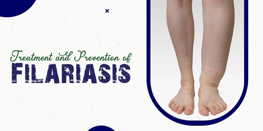
The chronic condition shows edema with thickening of the skin and underlying tissues (the classic symptom of filariasis).
It regularly impacts the lower extremities. However, the arms, vulva, breast, and scrotum (causing hydrocele creation) could also be infected. The edema in the extremities, chest, or genital area can result in the part becoming several times its average length due to blockage of the vessels of the lymphatic system.
Causes
Many cases of filariasis are generated by the parasite Wuchereria bancrofti. Culex, Aedes, and Anopheles mosquitoes serve as a vector for W. bancrofti in the transmission of the disease. Another parasite called Brugia malayi also causes the vector Mansonia and Anopheles mosquitoes to transmit filariasis.
Whenever an affected mosquito bites a nutritious person, the larvae, called microfilariae, transfer to the lymphatics and lymph nodes. Right here, they change into adult worms and might persist for several years.
The parasite, in turn, provides more microfilariae. The mosquitoes suck these microfilariae into the immaterial blood generally at night and during a bite. A similar sequence is then regular in one more healthy individual.
What is the prevention strategy?
Prevention involves giving a drug that kills the microscopic worms to the whole local community in the areas where the virus is prevalent. Skipping mosquito bites is the other method of prevention. These kinds of mosquitoes typically bite between dusk and dawn. You can carry out these actions if staying in an affected area: Sleep under a mosquito net. Use mosquito repellents on the exposed skin and take a yearly dose of a drug that kills the worms in the blood.